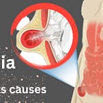
Definition Atheroma
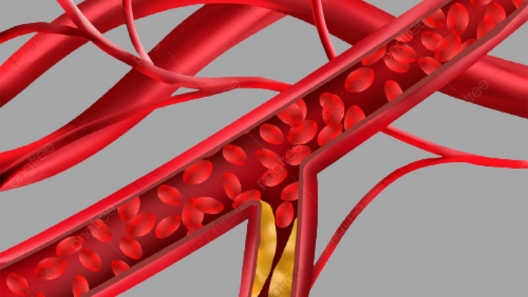
In this disease, the hard substance of the first color begins to accumulate inside the arteries,
due to which the arteries begin to narrow.
Sites of artery Atheroma
- In the arteries of the brain.
- In the arteries of the heart.
- In the Abdominal Aorta
- In the renal arteries.
- In The arteries are in the upper and lower parts of the body.
How does atheroma cause a heart attack
- This disease is more common due to obesity.
- Abundant consumption of meat and fat.
- Cigarettes, tea, coffee, alcohol in abundance.
- This disease is more common in men than in women.
- Due to high blood pressure.
- Due to living an unnatural life.
- Due to diabetes.
- This disease can also be hereditary.
What does atheroma effects
As we know that in this disease, the arteries are not narrowed and obviously, the blood is not
supplied to the body in its full amount from the narrowed arteries, due to which the following
things are seen:
- Myocar del infarction.
- Occurrence of necrosis.
Necrosis occurs when a part of the body is deprived of proper blood supply, turning blue
and cold, and eventually dies. That is, it decomposes. This condition is called “gangrene”
and the condition before decomposition “Negros” is called.
- Gangrene occurs.
- There is pain in the body. There is an excess of cholesterol in the blood.
Atheromatous plaque note
The fatty buildup in your arteries, whether you like to call it atheroma or plaque, isn’t helpful for
your health. Fighting atherosclerosis on your own, whether atheroma or plaque, is not effective
for your health. If you want to slow the growth of plaque in your arteries, discuss it with your
healthcare professional. Making lifestyle adjustments like giving up smoking and eating
well-balanced meals will help keep your arteries healthy for years to come.
Coronary artery atheroma formation
The process of atherosclerosis begins with issues in the endothelium that permit lipids
and cellular components to enter blood vessel walls. This leads to the build-up of lipids
in the arterial walls, which is the defining characteristic of this disease. To study this process,
researchers used DICOM images to create a computer model. They used Darcy’s law to model
the solid domain (arterial wall) and the Navier-Stokes equations with the continuity equation
to model the fluid domain (blood). By using convection-diffusion equations, low-density
lipoproteins (LDL) and oxygen were transported.
Calcified carotid atheroma on panoramic radiographs
Objective:
A stroke caused by carotid artery calcification accounts for approximately 5% of all strokes.
It is a common noninvasive procedure to evaluate carotid artery stenosis and calcification
with carotid Doppler ultrasonography (DS). In this study, we aimed to determine the level
of agreement between panoramic radiographs and DS results. Additionally, we sought to
evaluate the efficacy of panoramic radiography in detecting carotid artery calcification in
patients with and without coronary artery disease.
Methods:
It is estimated that about 5% of ischaemic strokes are related to carotid artery calcification.
Carotid Doppler (DS) ultrasonography is a commonly used noninvasive technique for evaluating
carotid artery stenosis and calcification. In this study, the level of agreement between
panoramic radiographs and DS findings was assessed as well as the ability of panoramic
radiography to detect carotid artery calcification in patients with and without
coronary artery disease.
Result:
The findings showed that panoramic radiography had a sensitivity of 66.6% and a positive
predictive value of 45% in detecting carotid artery calcifications in patients with coronary
artery disease. However, in patients with normal angiography, its sensitivity was 50%.
In addition, there was little agreement between the panoramic radiography results and
the DS results.
Stenotic coronary artery atheroma
In severe cases, osseous coronary artery stenosis may lead to death or reduced blood flow
from the distal coronary artery. Although atherosclerosis is the most common cause,
other factors such as aortic arteritis, congenital webs, and complications after aortic
valve replacement surgery can also contribute to this condition. During the autopsy,
three patients who died from ischemic heart disease were found to have an obscure ostial
stricture/occlusion for the first time. For example, in case 1, a 70-year-old man with right
coronary artery hypoplasia and a left anterior descending coronary artery tunnel had
atherosclerotically obliterated right coronary artery ostium.
It was observed in two cases of elderly males, ages 80 and 85, that they had considerable
epicardial coronary atherosclerosis, along with coronary obliteration in the right ostia.
In the case of sudden death, examining the coronary ostia is crucial as occlusive lesions
are often the sole cause of death. Additionally, ostial stenosis/occlusion can contribute
to global myocardial ischemia caused by coronary artery atheroma and/or other
factors in various situations.