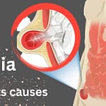

What is Embolism
Foreign objects can enter the bloodstream from both internal and external sources
and become trapped within the bloodstream.
How does Embolism cause stroke
- If air is present in the syringe while injecting, it can cause embolism as this air creates an airlock condition.
- As a result of a bone break, the bone pulp can enter the blood vessels and affect circulation, resulting in embolism.
- If the blood clot gets out of its place and joins the bloodstream, it is the cause of this disease.
- When the uterus opens during labor, multiple blood vessels allow air and amniotic fluid to enter. Amniotic Fluid This The fluid is in the membrane that wraps around the baby in the mother’s womb.
- A portion of a tumor can also embolize by entering the surrounding blood vessels.
Embolism Symptoms and Signs
- restlessness
- Air Hunger
- restlessness
- Shock may occur.
- pains.
A stroke may result from an embolism that affects the brain’s arteries.
-
Test
An angiography procedure is required to determine the problem.
Embolism Treatment Homeopathy Medicine
-
Arsenic album 30
Irritation, restlessness and anxiety, frequent thirst.
-
Chapter 30
There is pain and restlessness all over the body. other than that
C.Belladonna Arnica Brayta Carb Ammonia Lord Black Peacock Arithm
There is still value in Knight, etc.
Making the Diagnosis of Embolism
For the diagnosis of pulmonary emboli, there are numerous diagnostics available.
To further investigate if there are any obstructions in the blood flow to the lungs,
V/Q analysis, ventilation, and perfusion can be evaluated. Pulmonary angiography
and computed tomography are other options. When deep vein thrombosis occurs,
impedance plethysmography (IPG) of the veins and Doppler tests of the legs can
all be used to detect blood clots. In cases of strokes, where blood clots can block
arteries, brain scans, angiography, or Doppler ultrasounds may be performed.
Types of Embolism
- Pulmonary embolism: An embolism that occurs due to a traveling blood clot in the lungs
- Brain embolism: An embolism formed in the brain’s blood vessels
- Amniotic fluid embolism: a type of embolism caused by mixing amniotic fluid and fetal cells
- Air embolism: a blood clot caused by air or gas
- Fat embolism: An accumulation of fat particles can cause an embolism
- Septic embolism: An embolism caused by bacteria
- Retinal embolism: An embolism caused by blood clots or cholesterol blockages.
Pulmonary Embolism productive Cough
Thrombosis in the lungs occurs when a blood clot blocks a lung artery, preventing blood from
flowing through it. The Clot usually originates in a deep leg vein and travels to the lungs.
When a clot forms in one or more deep veins.
Pulmonary embolism can be life-threatening because it obstructs blood flow to the lungs.
It is significantly less likely that you will die if you receive prompt treatment. Prevent the
formation of blood clots in the legs by minimizing your risk of developing pulmonary
embolisms.
A cough that produces mucus or phlegm indicates something is wrong, while a dry cough
may not be as severe but can still be unpleasant and tiring. It also applies to the
cough that often accompanies moderate COVID-19. Some people may cough to
clear their throats, but others may cough frequently and even after eating or
consuming certain foods.
The causes of persistent dry cough vary and are unique to each individual.
Bronchoscopy Pulmonary Embolism
A procedure known as endobronchial ultrasonography is widely used to examine the
structures around the main airways. During this process, an ultrasonic probe at the
end of the bronchoscope is used to view and extract enlarged mediastinal lymph
nodes and masses. Furthermore, the aorta, main pulmonary artery, branches,
superior vena cava, and zygote vein can also be observed when searching for these lesions.
These can be further confirmed with an ultrasound processor in colour Doppler mode.
A thrombus or anomaly in these veins, such as an obstruction, can also be easily identified.
We discuss an unplanned but fascinating discovery made during one of these
observations in this article.
Flexible fiberoptic bronchoscopy with fine needle aspiration is currently used to diagnose
and evaluate lung diseases. However, this procedure has documented problems,
which are well-documented in the medical literature. Only four cases of air embolism
following fine needle aspiration during bronchoscopy have been reported. Despite the
many clinical symptoms associated with this issue, the clinical community has not fully
appreciated it. In the most recent British Thoracic Society statement on bronchoscopy
from 2013, this issue was not even mentioned. These two case reports describe incidents
of air embolism that occurred during a fine needle aspiration bronchoscopy.
Pulmonary Embolism in Puerperium
Pulmonary embolisms during pregnancy are becoming increasingly common.
The increase may be due to higher levels of high-risk populations, such as those
who have undergone reproductive technology or birth control, women who are
getting older, obese, and having cesarean deliveries, as well as better access to healthcare.
Pregnancy and after childbirth are often times not affected by venous thrombosis
and PE risk factors. In order to reduce maternal mortality, early diagnosis, prevention,
and prompt and effective treatment are essential. An angiography of the lungs,
ventilation and perfusion scans are common diagnostic tests. If necessary,
more severe treatments such as thrombolysis and effective anticoagulation are
also used, along with general supportive measures. Later health problems, such
as breastfeeding, contraception, mood swings, and recurrence in subsequent pregnancies,
should be addressed with postpartum treatment.
Conclusion
It is still one of the leading causes of maternal mortality in developed countries,
particularly among pregnant women, even though it is not very common.
In order to improve outcomes for both mother and child, it is imperative to
remain vigilant, diagnose the condition promptly, and treat it appropriately.
During and after pregnancy, even women who are initially at low risk can
become more susceptible. To fully understand these challenges,
further research is required.